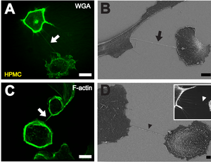
B Depiction of a TNT (black arrow) between two cells with scanning electron microscopy. Scale bar: 10 μm.
C Fluorescently labeled F-actin (white arrow) present in TNTs between individual HPMCs. Scale bar: 20 μm.
D Scanning electron microscope image of a potential TNT precursor (black arrowhead). Insert shows a fluorescence microscopic image of filopodia-like protrusions (white arrowhead) approaching a neighboring cell. Scale bar: 2 μm.[1]
A tunneling nanotube (TNT) or membrane nanotube is a term that has been applied to cytoskeletal protrusions that extend from the plasma membrane which enable different animal cells to connect over long distances, sometimes over 100 μm between certain types of cells.[2][3][4] Tunneling nanotubes that are less than 0.7 micrometers in diameter, have an actin structure and carry portions of plasma membrane between cells in both directions. Larger TNTs (>0.7 μm) contain an actin structure with microtubules and/or intermediate filaments, and can carry components such as vesicles and organelles between cells, including whole mitochondria.[5][6][7] The diameter of TNTs ranges from 0.05 μm to 1.5 μm and they can reach lengths of several cell diameters.[7][8] There have been two types of observed TNTs: open ended and closed ended. Open ended TNTs connect the cytoplasm of two cells. Closed ended TNTs do not have continuous cytoplasm as there is a gap junction cap that only allows small molecules and ions to flow between cells.[9] These structures have shown involvement in cell-to-cell communication, transfer of nucleic acids such as mRNA and miRNA between cells in culture or in a tissue, and the spread of pathogens or toxins such as HIV and prions.[10][11][12][13][14][3] TNTs have observed lifetimes ranging from a few minutes up to several hours, and several proteins have been implicated in their formation and inhibition, including many that interact with Arp2/3.[15][16]
- ^ Ranzinger J, Rustom A, Abel M, Leyh J, Kihm L, Witkowski M, et al. (2011-12-27). Bereswill S (ed.). "Nanotube action between human mesothelial cells reveals novel aspects of inflammatory responses". PLOS ONE. 6 (12): e29537. Bibcode:2011PLoSO...629537R. doi:10.1371/journal.pone.0029537. PMC 3246504. PMID 22216308.
- ^ Abounit S, Zurzolo C (March 2012). "Wiring through tunneling nanotubes--from electrical signals to organelle transfer". Journal of Cell Science. 125 (Pt 5): 1089–1098. doi:10.1242/jcs.083279. PMID 22399801. S2CID 8433589.
- ^ a b Sowinski S, Jolly C, Berninghausen O, Purbhoo MA, Chauveau A, Köhler K, et al. (February 2008). "Membrane nanotubes physically connect T cells over long distances presenting a novel route for HIV-1 transmission". Nature Cell Biology. 10 (2): 211–219. doi:10.1038/ncb1682. PMID 18193035. S2CID 25410308.
- ^ Davis DM, Sowinski S (June 2008). "Membrane nanotubes: dynamic long-distance connections between animal cells". Nature Reviews. Molecular Cell Biology. 9 (6): 431–436. doi:10.1038/nrm2399. PMID 18431401. S2CID 8136865.
- ^ Resnik N, Erman A, Veranič P, Kreft ME (October 2019). "Triple labelling of actin filaments, intermediate filaments and microtubules for broad application in cell biology: uncovering the cytoskeletal composition in tunneling nanotubes". Histochemistry and Cell Biology. 152 (4): 311–317. doi:10.1007/s00418-019-01806-3. PMID 31392410. S2CID 199491883.
- ^ Onfelt B, Nedvetzki S, Benninger RK, Purbhoo MA, Sowinski S, Hume AN, et al. (December 2006). "Structurally distinct membrane nanotubes between human macrophages support long-distance vesicular traffic or surfing of bacteria". Journal of Immunology. 177 (12): 8476–8483. doi:10.4049/jimmunol.177.12.8476. PMID 17142745.
- ^ a b Rustom A, Saffrich R, Markovic I, Walther P, Gerdes HH (February 2004). "Nanotubular highways for intercellular organelle transport". Science. 303 (5660): 1007–1010. Bibcode:2004Sci...303.1007R. doi:10.1126/science.1093133. PMID 14963329. S2CID 37863055.
- ^ Wang ZG, Liu SL, Tian ZQ, Zhang ZL, Tang HW, Pang DW (November 2012). "Myosin-driven intercellular transportation of wheat germ agglutinin mediated by membrane nanotubes between human lung cancer cells". ACS Nano. 6 (11): 10033–10041. doi:10.1021/nn303729r. PMID 23102457.
- ^ Zurzolo C (August 2021). "Tunneling nanotubes: Reshaping connectivity". Current Opinion in Cell Biology. Membrane Trafficking. 71: 139–147. doi:10.1016/j.ceb.2021.03.003. PMID 33866130. S2CID 233298036.
- ^ Onfelt B, Davis DM (November 2004). "Can membrane nanotubes facilitate communication between immune cells?". Biochemical Society Transactions. 32 (Pt 5): 676–678. doi:10.1042/BST0320676. PMID 15493985. S2CID 32181738.
- ^ Haimovich G, Dasgupta S, Gerst JE (February 2021). "RNA transfer through tunneling nanotubes". Biochemical Society Transactions. 49 (1): 145–160. doi:10.1042/BST20200113. PMID 33367488. S2CID 229689880.
- ^ Haimovich G, Ecker CM, Dunagin MC, Eggan E, Raj A, Gerst JE, Singer RH (November 2017). "Intercellular mRNA trafficking via membrane nanotube-like extensions in mammalian cells". Proceedings of the National Academy of Sciences of the United States of America. 114 (46): E9873–E9882. Bibcode:2017PNAS..114E9873H. doi:10.1073/pnas.1706365114. PMC 5699038. PMID 29078295.
- ^ Belting M, Wittrup A (December 2008). "Nanotubes, exosomes, and nucleic acid-binding peptides provide novel mechanisms of intercellular communication in eukaryotic cells: implications in health and disease". The Journal of Cell Biology. 183 (7): 1187–1191. doi:10.1083/jcb.200810038. PMC 2606965. PMID 19103810.
- ^ Gousset K, Schiff E, Langevin C, Marijanovic Z, Caputo A, Browman DT, et al. (March 2009). "Prions hijack tunnelling nanotubes for intercellular spread". Nature Cell Biology. 11 (3): 328–336. doi:10.1038/ncb1841. PMID 19198598. S2CID 30793469.
- ^ Gurke S, Barroso JF, Gerdes HH (May 2008). "The art of cellular communication: tunneling nanotubes bridge the divide". Histochemistry and Cell Biology. 129 (5): 539–550. doi:10.1007/s00418-008-0412-0. PMC 2323029. PMID 18386044.
- ^ Hanna SJ, McCoy-Simandle K, Miskolci V, Guo P, Cammer M, Hodgson L, Cox D (August 2017). "The Role of Rho-GTPases and actin polymerization during Macrophage Tunneling Nanotube Biogenesis". Scientific Reports. 7 (1): 8547. Bibcode:2017NatSR...7.8547H. doi:10.1038/s41598-017-08950-7. PMC 5561213. PMID 28819224.