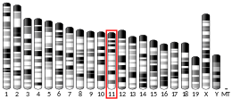
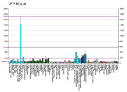
Voltage-dependent anion-selective channel 1 (VDAC-1) is a beta barrel protein that in humans is encoded by the VDAC1 gene located on chromosome 5.[4][5] It forms an ion channel in the outer mitochondrial membrane (OMM) and also the outer cell membrane. In the OMM, it allows ATP to diffuse out of the mitochondria into the cytoplasm. In the cell membrane, it is involved in volume regulation. Within all eukaryotic cells, mitochondria are responsible for synthesis of ATP among other metabolite needed for cell survival. VDAC1 therefore allows for communication between the mitochondrion and the cell mediating the balance between cell metabolism and cell death. Besides metabolic permeation, VDAC1 also acts as a scaffold for proteins such as hexokinase that can in turn regulate metabolism.[6]
This protein is a voltage-dependent anion channel and shares high structural homology with the other VDAC isoforms (VDAC2 and VDAC3), which are involved in the regulation of cell metabolism, mitochondrial apoptosis, and spermatogenesis.[7][8][9][10] Over expression and misregulation of this pore could lead to apoptosis in the cell leading to a variety of diseases within the body. In particular, since VDAC1 is the major calcium ion transport channel, its dysfunction is implicated in cancer, Parkinson's (PD), and Alzheimer's disease.[11][12][13] In addition, recent studies have shown that an over expression within the VDAC1 protein is linked to Type 2 Diabetes. Lund University released a study that demonstrated the effects of blocking VDAC1 over expression can prevent the spread of Type 2 Diabetes.[14]

- ^ a b c GRCm38: Ensembl release 89: ENSMUSG00000020402 – Ensembl, May 2017
- ^ "Human PubMed Reference:". National Center for Biotechnology Information, U.S. National Library of Medicine.
- ^ "Mouse PubMed Reference:". National Center for Biotechnology Information, U.S. National Library of Medicine.
- ^ Blachly-Dyson E, Baldini A, Litt M, McCabe ER, Forte M (March 1994). "Human genes encoding the voltage-dependent anion channel (VDAC) of the outer mitochondrial membrane: mapping and identification of two new isoforms". Genomics. 20 (1): 62–67. doi:10.1006/geno.1994.1127. PMID 7517385.
- ^ "Entrez Gene: VDAC1 voltage-dependent anion channel 1".
- ^ Cite error: The named reference
pmid20434446was invoked but never defined (see the help page). - ^ Subedi KP, Kim JC, Kang M, Son MJ, Kim YS, Woo SH (February 2011). "Voltage-dependent anion channel 2 modulates resting Ca²+ sparks, but not action potential-induced Ca²+ signaling in cardiac myocytes". Cell Calcium. 49 (2): 136–143. doi:10.1016/j.ceca.2010.12.004. PMID 21241999.
- ^ Alvira CM, Umesh A, Husted C, Ying L, Hou Y, Lyu SC, et al. (November 2012). "Voltage-dependent anion channel-2 interaction with nitric oxide synthase enhances pulmonary artery endothelial cell nitric oxide production". American Journal of Respiratory Cell and Molecular Biology. 47 (5): 669–678. doi:10.1165/rcmb.2011-0436OC. PMC 3547107. PMID 22842492.
- ^ Cheng EH, Sheiko TV, Fisher JK, Craigen WJ, Korsmeyer SJ (July 2003). "VDAC2 inhibits BAK activation and mitochondrial apoptosis". Science. 301 (5632): 513–517. Bibcode:2003Sci...301..513C. doi:10.1126/science.1083995. PMID 12881569. S2CID 37099525.
- ^ Li Z, Wang Y, Xue Y, Li X, Cao H, Zheng SJ (February 2012). "Critical role for voltage-dependent anion channel 2 in infectious bursal disease virus-induced apoptosis in host cells via interaction with VP5". Journal of Virology. 86 (3): 1328–1338. doi:10.1128/JVI.06104-11. PMC 3264341. PMID 22114330.
- ^ Huang H, Shah K, Bradbury NA, Li C, White C (October 2014). "Mcl-1 promotes lung cancer cell migration by directly interacting with VDAC to increase mitochondrial Ca2+ uptake and reactive oxygen species generation". Cell Death & Disease. 5 (10): e1482. doi:10.1038/cddis.2014.419. PMC 4237246. PMID 25341036.
- ^ Chu Y, Goldman JG, Kelly L, He Y, Waliczek T, Kordower JH (September 2014). "Abnormal alpha-synuclein reduces nigral voltage-dependent anion channel 1 in sporadic and experimental Parkinson's disease". Neurobiology of Disease. 69: 1–14. doi:10.1016/j.nbd.2014.05.003. PMID 24825319. S2CID 22722682.
- ^ Smilansky A, Dangoor L, Nakdimon I, Ben-Hail D, Mizrachi D, Shoshan-Barmatz V (December 2015). "The Voltage-dependent Anion Channel 1 Mediates Amyloid β Toxicity and Represents a Potential Target for Alzheimer Disease Therapy". The Journal of Biological Chemistry. 290 (52): 30670–30683. doi:10.1074/jbc.M115.691493. PMC 4692199. PMID 26542804.
- ^ Zhang E, Mohammed Al-Amily I, Mohammed S, Luan C, Asplund O, Ahmed M, et al. (January 2019). "Preserving Insulin Secretion in Diabetes by Inhibiting VDAC1 Overexpression and Surface Translocation in β Cells". Cell Metabolism. 29 (1): 64–77.e6. doi:10.1016/j.cmet.2018.09.008. PMC 6331340. PMID 30293774.


