| Ventricular septal defect | |
|---|---|
 | |
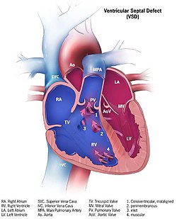
| Illustration showing various forms of ventricular septal defects. 1. Conoventricular, malaligned 2. Perimembranous 3. Inlet 4. Muscular | |
| Specialty | Cardiac surgery |
A ventricular septal defect (VSD) is a defect in the ventricular septum, the wall dividing the left and right ventricles of the heart. The extent of the opening may vary from pin size to complete absence of the ventricular septum, creating one common ventricle. The ventricular septum consists of an inferior muscular and superior membranous portion and is extensively innervated with conducting cardiomyocytes.
The membranous portion, which is close to the atrioventricular node, is most commonly affected in adults and older children in the United States.[1] It is also the type that will most commonly require surgical intervention, comprising over 80% of cases.[2]
Membranous ventricular septal defects are more common than muscular ventricular septal defects, and are the most common congenital cardiac anomaly.[3]
- ^ Taylor, Michael D (2019-02-02). "Muscular Ventricular Septal Defect". EMedicine. Medscape.
- ^ Waight, David J.; Bacha, Emile A.; Kahana, Madelyn; Cao, Qi-Ling; Heitschmidt, Mary; Hijazi, Ziyad M. (March 2002). "Catheter therapy of Swiss cheese ventricular septal defects using the Amplatzer muscular VSD occluder". Catheterization and Cardiovascular Interventions. 55 (3): 355–361. doi:10.1002/ccd.10124. PMID 11870941. S2CID 23602868.
- ^ Hoffman, JI; Kaplan, S (2002). "The incidence of congenital heart disease". Journal of the American College of Cardiology. 39 (12): 1890–900. doi:10.1016/S0735-1097(02)01886-7. PMID 12084585.